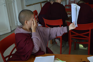
When asked what they saw, the nuns all said that the image was upside down and reversed left and right. As it should be. This is similar to the image that is projected onto the retina; the brain “flips” the image right-side up.
We continued to explore the retina in more detail by discussing photoreceptors (rods and cones). To determine the distribution of rods and cones on the retina, the nuns drew a protractor on a piece of paper and labeled it with degrees. To create a tester, the nuns attached small, colored numbers to a stick. With one nun looking straight ahead, the other nun moved the tester from peripheral vision to central vision. The nuns were asked to record when (what degree) they could first see movement, color and detail. As expected, movement could be detected in the periphery, but color and detail required more central vision. This experiment demonstrated how cones (which respond to different frequencies of light) are found in greatest numbers in more central parts of the retina.
I also mentioned that there is one small spot in each retina here there are no photoreceptors. This is our blind spot: the region of the retina where axons from ganglion cells exit the eye on their way to the brain. We made several types of blind spot testers to see how the brain fills in the visual gap created by the blind spot.
We ended our discussion of vision by talking about monocular and binocular vision. Why do we have two eyes instead of one? What is the advantage of having two eyes? We played a game to find out. Each nun was given a bead and asked to throw it into a dish using two eyes and then again using one eye. Many more beads ended up in the dish with they use two eyes indicating that two eyes allow for better depth perception.


No comments:
Post a Comment